- Recent Projects
- Machine learning enabled cell imaging
- oldmasterprojects
- Air coupled ultrasonic inspection of thin walled plates
- Electrical transport in graphene nanoribbons bridging carbon nanotube gaps
- Spatially mapping thermal conductivity of 2D-materials
- Electrical and optical biosensors
- B
- H2020 project MOTIVATE [Link to other Website]
- Publications 2017
- News-List2
- Processes
- hiddentestpage2
- hiddentestpage
- Testpage
- Team2
- Measurement Technology
- Tech Transfer & Services
- Education
- All Publications R
- News List
- Development of microporous graphene membranes
- Publications 2017 Ref
- Publications 2016 Ref
- Publications 2015 Ref
- All Publications
- Oxide based resistive switching memories for neuromorphic computing
- THz Talbot imaging
- THz tomography
- Publications 2016 2
- Opto-electronic characterization of strain effects in graphene nanoribbons
- Artificial Intelligence for the analytics of quantum devices
- Silicon Nanowires for sensing
- Equipment
- Scanning Thermal Microscopy of suspended graphene
- Reliability and safety
- Publication extract (internal)
- Current Projects
- news-list3
- Welcome1
- DemoListRight
- Welcome2
- logfile
- Empa-Dora-testpage
Machine learning enabled cell imaging
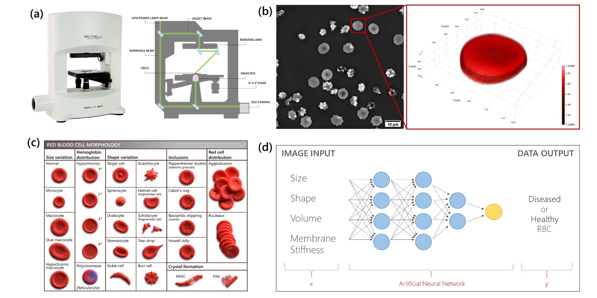
Red Blood Cells (RBCs) are the most abundant cell type in blood, with their primary function being the transport of oxygen throughout the body. RBCs extracted from donors diagnosed with pathological diseases tend to exhibit morphological abnormalities, which can be measured and characterized. The goal of this project is to develop an unconventional imaging platform for the reliable and rapid fingerprinting of pathological diseases by monitoring the shape, size and morphological changes occurring in RBCs. Our unique approach relies upon the combination of high-throughput live cell imaging with high resolution scanning electron microscopy and scanning probe microscopy methodologies. The data obtained from these state-of-the-art techniques will be collected and analyzed using Machine Learning (ML) algorithms. ML-based data analytics will provide an automated and more standardized approach for RBC classification in health and blood related diseases, with the goal of supporting clinicians in improving early detection, prognosis and the development of more effective treatment strategies. This project strives for the realization of digital pathology and its implementation in the advancements of precision medicine.


