Seeing blood at the nanoscale
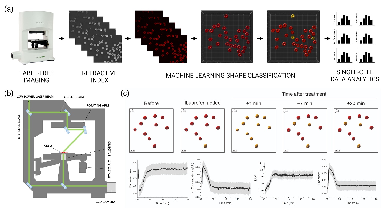
Blood is the most complex biofluid in our body and it consists of different circulating cell types, including red blood cells, white blood cells and platelets, and blood prod-ucts, such as blood clots. Investigating the morphology and biophysical functions of blood components is crucial for uncovering valuable disease biomarkers and for the monitoring of side effects induced by drugs (e.g. ibuprofen) or disease. To this end, we use digital holo-tomographic microscopy, scanning probe microscopy and machine learning-based image and data analysis to characterize and structurally classify blood components in health and disease, without the need for laborious and cell harming staining procedures. Within this research work, we focus on investigating the real-time dose-dependent chemical effects on red blood cells and blood clots, with the ultimate goal of supporting clinicians in improving early disease detection, prognosis and in the development of more effective and personalized treatment strategies.


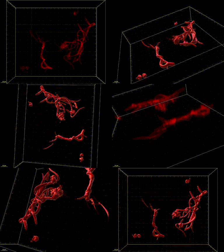
(a) Label-free imaging and machine learning-based image analysis framework used within this research project. (b) Schematic and working principle of digital holo-tomographic mi-croscopy (DHTM, 3D Cell Explorer, Nanolive SA) for quantitative, non-invasive and label-free 3D live cell imaging. (c) Representative time-lapse images before and after treatment with ibuprofen (red = normocyte, yellow = echinocyte) and quantitative representation of time-dependent biologically-relevant morphological parameters, including cell diameter, hemoglobin concentration, surface-area-to-volume (SA:V) ratio and sphericity. GIF: 3D re-fractive index reconstruction of a blood clot fragment imaged using DHTM.
Publications
Bergaglio, T., Bhattacharya, S., Thompson, D., & Nirmalraj, P. N.
ACS Nanoscience Au. (2023).


