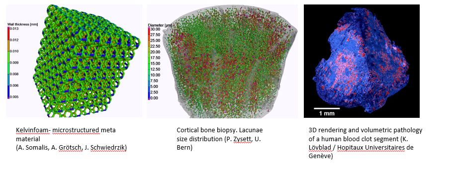
Low-energy X-ray Imaging
The development of innovative and novel X-ray Computed-Tomography (XCT) methods is an outstanding capability of Empa's Center for X-ray Analytics. Combined with state-of-the-art image reconstruction and processing, using in-house and commercial tools, it offers quantitative and qualitative non-destructive analysis for a wide range of materials and samples with spatial resolutions down to sub-micrometer levels.
Dedicated X-ray imaging methods are available for specific materials. Low-attenuation-contrast materials can be analysed using different X-ray Phase Contrast Imaging (PCI) methods including propagation-based, speckle-based methods and grating interferometry. For samples with nano-structures X-ray Dark-Field Imaging (DFI) tools using speckle-based methods and grating interferometry are utilised. This extensive know-how is complemented with development projects for novel X-ray sources and detectors. The Center offers unique possibilities to advance X-ray analytics to new levels and make it usable for classes of materials where it was not available before.
Basic and applied developments in the domains of bio- and life-sciences, personalized medicine, coating characterisation, wood analysis, micro/nano-structured scaffolds, additive manufacturing, concrete and asphalt structures, bone-mineral density analysis, biofilm imaging, cartilage imaging, artefact reduction and many others are part of the activities of the group.


