3D Image Analysis and Simulation
The main goals in this area are
- providing users of the X-ray analytics/imaging facilities at Empa computational methods and tools for the quantitative analysis of their datasets, with specific focus on tomographic datasets;
- supporting the development of new analytics/imaging experimental methods by developing/implementing signal/image processing algorithms based upon the theoretical/computational analysis of the physical processes involved in the image formation.
To achieve these goals
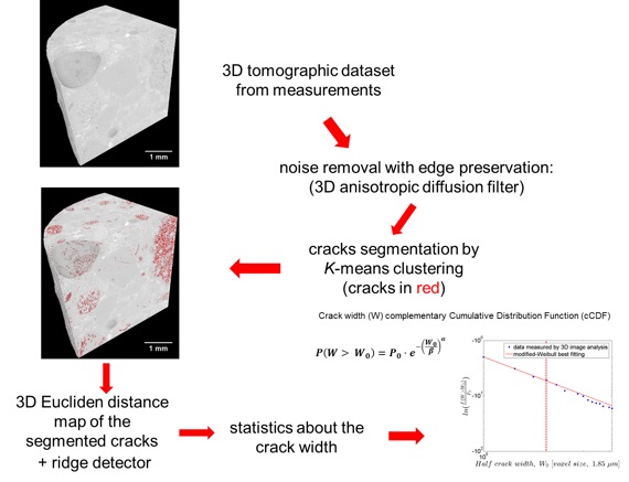
- we perform research & development in the field of 3D image analysis, including the development of new analysis algorithms and workflows, customized for specific tasks and types of datasets/materials, and the algorithm implementation into in-house developed software; see Fig. 1 for an example of a 3D image analysis workflow.
- we perform research & development on tomographic reconstruction algorithms for reducing beam hardening, metal and ring artifacts in the final images and for multi-contrast (see the X-ray Phase Contrast Imaging area) and fast X-ray imaging, the latter to be used in dynamic tomography or tomography during in situ experiments; the algorithms are implemented in in house-developed software exploiting advanced high performance computing (HPC) methods, e.g., GPU processing. The reconstruction software is free available as open source (https://github.com/JueHo/CT-Recon).
- we provide the users of the facilities at Empa support with their tomographic reconstruction, image processing/analysis needs and access to HPC software/hardware resources for fulfilling them; see the Empa Platform for Image Analysis info page.
- we promote the development of an imaging/image analysis user community, by the organization of Topical Days on these topics at the Empa Academy.

Example of 3D image analysis workflow for a very high spatial resolution X-ray tomographic dataset, acquired during measurements at the TOMCAT beamline of the Swiss Light Source (PSI). The material is concrete. The sample was a cylinder, about 7 mm in diameter and 40 mm long. A region of interest (ROI) was imaged, 3.256 mm long along the cylinder symmetry axis. Only sub-ROI is rendered in this figure. The concrete sample was subjected to a laboratory-scale protocol for accelerating a set of chemical reactions (alkali-aggregate reaction, AAR) occurring with a very slow kinetics (tens of years) in real-world concrete structures. The AAR leads to cracking. 3D image analysis, with algorithms developed and implemented at Empa, allows extracting quantitative information about the cracking geometrical, morphological and topological properties, after having identified the voxels in the dataset corresponding to cracks (in red).
The obtained quantitative information is used for both basic research purposes, e.g., validation of computational models of AAR-induced cracking, and practical applications, e.g., non-destructive, digital analysis of cores from laboratory-scale samples used in experimental studies or from samples coming from actual concrete structure affected by this type of damage. In the example shown here, the quantitative information extracted from the tomographic datasets is the complementary cumulative distribution function (cCDF) for the crack width, expressed in voxel size units (consider only the range above 2). The tomography-derived data are compared with an analytical model for the cCDF (best fitting by a Weibull cCDF).


-
Share
