Instrumentation
Empa's Electron Microscopy Center operates a modern infrastructure of electron microscopy equipment for materials and nano-science. Besides scanning electron microscopes and (scanning) transmission electron microscopes equipped with analytical devices, the center maintains facilities for sample preparation of solid (nano-)materials. For advanced data analysis various software packages, including software for simulations are available.
Transmission Electron Microscopes
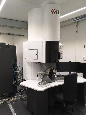
FEI Titan Themis
The Titan Themis, operational at 80 and 300 kV, provides highest resolution imaging capabilties for analytics and particularly for in-situ and in-operando measuremetns. The microscope is equipped with a hexapole-type aberration corrector for scanning transmission electron microscope (CEOS DCOR) and a SuperEDX system. In situ sample holders from Protochips can be used for liquid cell measurements and thermo-electrical measurements. A fast gun valve provides additional safety for in situ experiments.
Main features & techniques:
- High-resolution scanning transmission electron microscopy (<60 pm)
- SuperEDX system
- CEFID EELS / EFTEM system
- Differential phase contrast imaging (DPC and iDPC)
- Tomography (STEM & TEM)
- Electrostatic biprism for electron holography
- Lorentz lens
- FEI CETA 2 camera (16 Mpixel, up to 40 fps at full resolution)
- Protochips Poseidon holder (dual flow and heating)
- Protochips Fusion 500 holder (double-tilt)
Installed: 2016
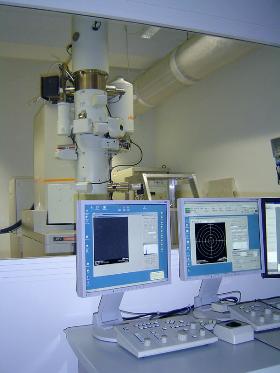
JEOL JEM2200fs
The JEOL JEM 2200fs (scanning) transmission electron microscope is an analytical multifunctional microscope equipped with a Schottky field-emission electron source. It features an Omega-type electron energy filter, an energy-dispersive X-ray spectrometer, a scanning unit and a variety of different sample holders.
Main features & techniques:
- High-resolution imaging in TEM and STEM mode (~2 Å resolution)
- Analytics: EELS, EFTEM & EDX
- Tomography
- Holders: tomography, liquid-nitrogen, heating, quartet, Be-single and Be-double tilt
- Gatan DigiScan II
Installed: 2009
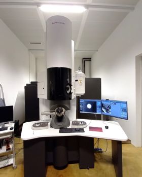
Titan Themis 200 G3
The Thermo Scientific Titan Themis 200 G3 offers advanced TEM, STEM and electron diffraction capabilities. Outfitted with a state-of-the-art probe corrector, a resolution of <80 pm can be reached in STEM mode. A SuperX detector allows for spatially resolved EDX mappings down to the atomic scale. Further capabilities of the instrument include high-speed TEM movie acquisition, tomography and in situ heating.
Main features & techniques:
JEOL ARM200F
Owned by and installed at IBM Zurich, staff of Empa's Electron Microscopy Center jointly operates this double-aberration-corrected (scanning) transmission electron microscope with a team of IBM scientists. The microscope is installed in a noise-free lab of the Binning-Rohrer Nanotechnology Center and is equipped with a cold field-emission electron source, a high-efficiency EDX detector, a electron energy-loss spectrometer and two correctors for the spherical aberration, one for the scanning transmission mode and one for the broad-beam transmission mode.
Main features & techniques:
- High-resolution imaging in TEM and STEM mode (<1 Å resolution)
- Operational at 80, 120 and 200 kV
- Analytics: EELS & EDX
- Gatan DigiScan II
Installed: 2014
Scanning Electron Microscopes
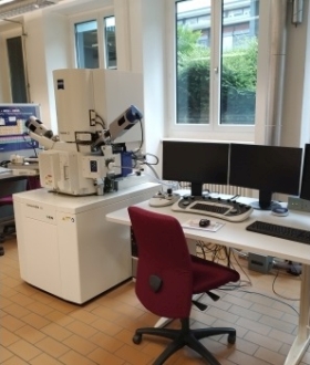
ZEISS GeminiSEM 460
The Zeiss GeminiSEM 460 is a multifunctional field emission scanning electron microscope with a double condenser system for high-resolution imaging and high-efficiency analytics. The ZEISS GeminiSEM 460 is jointly owned and operated with EAWAG.
Main features:
- High vacuum mode (HV: <10-2 Pa), standard variable pressure mode (VP: 5 - 60 Pa) and enhanced VP mode (NanoVP: 5 - 40 Pa)
-
Variety of detectors for signal optimization (SE, Inlens-SE, BSD and EsB detector)
-
Oxford EDS system (Ulitm Max 170 and Ulitm Extreme)
-
Oxford EBSD system (Symmetry S2)
-
STEM mode
Installed: 2022
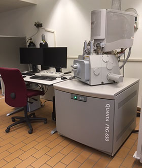
FEI Quanta 650 FEG ESEM
The FEI Quanta 650 is a multifunctional field-emission scanning electron microscopes with environmental SEM capabilities. It features:
- High-vacuum, low-vacuum and environmental (<26 mbar H2O) modi
- Peltier/heating sample stage
- Thermo Scientific Noran System 7 (UltraDry EDS Detector)
- Tensile stage
- Variety of detectors for signal optimization
Installed: 2017
Sample Preparation
- Ion mill Fischione Model 1050 TEM Mill
- Plasma cleaner Fischione 1020
- Dimple grinder
- Tripod polisher
- Manual mechanical polisher Struers
- Automatic mechanical polisher: Allied MultiPrep 8"
- Microtome Ultracut
- Sputterer Leica ACE600
- Carbon coater Safematic CCU-010
- Electropolisher Struers TenuPol-5
- Diamond-wire saw
Data Analysis and Simulations
Aside from various computer codes developed by staff of the Electron Microscopy Center, which is used for the analysis and simulation for in-line electron holography, scanning transmission electron micrographs and low-loss electron energy-loss spectroscopy, the following commercially available computer programs are available:
- Simulation and data analysis: MacTempas & CrystalKit from Total Resolution
- Simulations (TEM, STEM, Diffraction): xHREM from HREM Research
- Simulation (TEM, STEM, Diffraction): JEMS
- EELS simulation: Wien2K, FEFF8
- First-principles programs for EELS simulations: Wien2k & FEFF8
- Data analysis: Geometrical Phase Analysis and SmartAlign from HREM Research
- Tomographic reconstruction: Inspect 3D from FEI and TomoJ running on ImageJ
- 3D visualization and volume analysis: Amira
- Focal series reconstruction: TrueImage from FEI and MacTempas from Total Resolution
- Other software tools for microscopy data: ImageJ, Gatan Digital Micrograph, FEI TEM Image & Analysis (TIA), FEI Velox, Bruker ESPRIT, Oxford INCA
- Various "home"-written skripts and programs are used for data analysis and simulations (based on Fortran77, Mathematica, MATLAB etc.)
