Diagnostics
Detecting Alzheimer's in blood
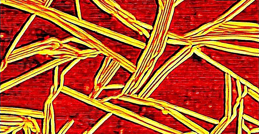
Empa researcher Peter Nirmalraj wants to capture images of Alzheimer's peptides with unprecedented precision. This could allow new insights into the molecular pathogenesis of the neurodegenerative disease – and perhaps open the way to new therapies.

In the beginning, physicist Peter Nirmalraj wanted to gain deeper insights into the process of Alzheimer's disease in order to provide medical research with a basis for possible early diagnostics and new therapies. One step further would be to decipher the role of amyloid beta peptides and tau proteins, which are associated with the neurodegenerative disease.
However, if one approaches the tiny peptide structures with a microscope, the view into the molecular universe becomes distorted all too easily. "An imaging technique must not alter the original state of the peptides, otherwise valuable information on their characteristic properties will be lost" Nirmalraj says. That's why the Empa scientist searched for a technique that left the peptides pristine.
Thanks to an ultra-clean, pre-treated graphene surface, he was able to capture images of the beta amyloid peptides with an atomic force microscope (AFM) without damaging their natural size, shape and morphology. In addition to the amyloid fibers, even smaller peptide particles, known as oligomers, can be seen. Their role in the pathogenic process may be more important than previously thought, suspects Nirmalraj. Currently, the lab results are being taken further in a pilot study with a team at the Cantonal Hospital of St. Gallen. The study aims to show if and how AFM technology can detect amyloid and tau proteins in blood samples from Alzheimer's patients.
|
Further information Dr. Peter Nirmalraj |
Editor / Media contact |
Diagnostics
Early detection of dementia
Alzheimer's and other dementias are among the most widespread diseases today. Diagnosis is complex and can often only be established with certainty late in the course of the disease. A team of Empa researchers, together with clinical partners, is now developing a new diagnostic tool that can detect the first signs of neurodegenerative changes using a sensor belt.
>>>>

Fibers
A data river runs through it
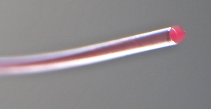
Data and signals can be transmitted quickly and reliably with glass fibers – as long as the fiber does not break. Strong bending or tensile stress can quickly destroy it. An Empa team has now developed a fiber with a liquid glycerol core that is much more robust and can transmit data just as reliably. And such fibers can even be used to build microhydraulic components and light sensors.
>>>>

Gloss
RIP, Hypatia …
Hypatia, the joint cluster computer of Empa and Eawag, is no more. On 10 March, the well-behaved and fast helper for scientific simulations was dismantled in Empa's basement and loaded onto the trucks of a waste disposal company. Many components of the mainframe will now begin a new life elsewhere. Hypatia was named after one of the few female scientists of antiquity who lived in Egypt in the 4th century.
>>>>
