Translational Musculoskeletal Biodynamics
Our primary goal is to uncover critical biomechanical markers and mechanical profiles that are unique to degenerative musculoskeletal diseases. To accomplish this, we combine in vivo multi-modality imaging with expertise in biomechanics, clinical and preclinical imaging, material science and big data management. Within this larger context, our current focus is on clinically relevant orthopedic biomechanical aspects of musculoskeletal disorders.

Musculoskeletal disorders are the second most common cause of disability worldwide. Understanding the dynamic relationship between mechanical forces and biochemical signaling pathways is one the major challenges in orthopedics.
Despite movement being a fundamental aspect of the human musculoskeletal system, traditional imaging techniques such as static X-rays, computed tomography (CT) and magnetic resonance imaging (MRI) require patients to remain still, limiting their ability to capture dynamic conditions.
Our long-term goal is to fill the gap with respect to identification of reliable biomarkers and detailed characterization of disorder phenotypes of degenerative musculoskeletal joint disorders from multi- modality in vivo imaging, and exploit this knowledge to enable appropriate, patient-specific diagnoses and treatment strategies. In this regard, dynamic assessment of in vivo three-dimensional (3D) joint kinematics, particularly during functional activities is essential for understanding normal joint function, and, correspondingly, the aberrations due to injury or disease. Ideally, a kinematic data acquisition set up should afford the capability of directly recording continuous, 3D motion of each of the bones comprising the joint in question, preferably in vivo and during functional activities, to capture the gross rotational movements such as flexion, extension, abduction and adduction—osteo-kinematics—, but also the finer rolling and sliding movements—arthro-kinematics with high fidelity and accuracy.
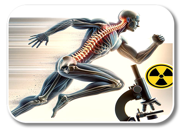
To address this, we utilize highly customized, high-speed dynamic biplanar radiographic imaging (DBRI) in combination with conventional skin-marker based video motion capture (MoCap) systems, as available in a traditional gait analysis laboratory. These moving X-ray images, along with simultaneously recorded datasets from MoCap, ground reaction forces and muscle electromyography (EMG), complement information gleaned from traditional three-dimensional imaging techniques like MRI and CT, offering a more comprehensive view of joint behavior and movement patterns. By combining these imaging modalities, we gain valuable insights into conditions like osteoarthritis, back pain and joint instability, improving diagnostic accuracy and enabling patient-specific treatments.
Related Projects – additional Images and References to be added
