
Infrastructure & Imaging
The Particles-Biology lab has access to state-of-the-art research and development laboratory rooms at Empa with modern infrastructure, specialized equipment and the long-standing expertise of our in-house specialists for material synthesis, material characterization and various biosafety level 2 in vitro and ex vivo human-based models.
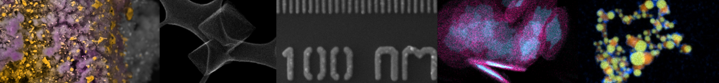
Imaging
In order to study different materials (see center of scheme below) and how they interact with different biosystems (right side of scheme), such as human cell-based samples, microbes or fungi (left side of scheme), we heavily rely on our advanced in-house live, in situ and correlative imaging capabilities.
We use state-of-the-art light microscopy (LM), confocal laser scanning microscopy (CLSM) and various transmission and scanning electron microscopy techniques ((cryo-)TEM, SEM, HR-(S)TEM, tomography, ultramicrotomy) for our research.
Additionally, we develop novel sample preparation, electron microscopy, imaging and correlative workflows to suit our broad array of research questions and material/biological samples. Moreover, we collaborate and are open to research collaborations with Swiss and international universities, research institutions, hospitals and industry partners.

Analytics
Complementing our imaging expertise and embedded in our correlative and complementary workflows we employ several analytical techniques that give us insight into element content, - speciation and - distribution as well as (particle) size, morphology and structure. These include techniques available in our own labs such as elemental analysis via Inductively Coupled Plasma Mass or Optical Emission Spectrometry ((sp)ICP-MS/ICP-OES) and SEM-coupled Energy Dispersive X-ray Spectroscopy (EDX) as well as techniques available from Empa-partners (X-ray photoelectron spectroscopy (XPS), confocal Raman microscopy, and X-ray diffraction (XRD)) and the ETH domain (high-resolution STEM-EDX and electron energy loss spectroscopy (EELS) at ScopeM and field-flow fractionation coupled to dynamic and/or static light scattering at Eawag).
An important consideration in developing standards and regulations that govern the production and use of commercial nanoscale materials is the development of robust and reliable measurements to estimate with high confidence the physical-chemical properties as well as the potential adverse biological effects.
In the H2020 EU-NCL and H2020 Refine projects we developed protocols for nanoparticles size distribution, zeta potential measurement and in vitro assays (e.g. cell viability) within a global network (NIST, JRC, KRISS and Nanotech Thailand). These concepts and protocols are published (see publications).

In the new HEurope project Metrino (Metrology for innovative nanotherapeutics) we are developing AI-based image analysis routines for (correlative) electron microscopy to advance the development of measurement methods to detect, localize and quantify NPs in biological tissues, to compare the results obtained by orthogonal techniques, and to evaluate their robustness and associated uncertainty.


