3D Micro-CT virtual histopathology for precision diagnostics for tumors
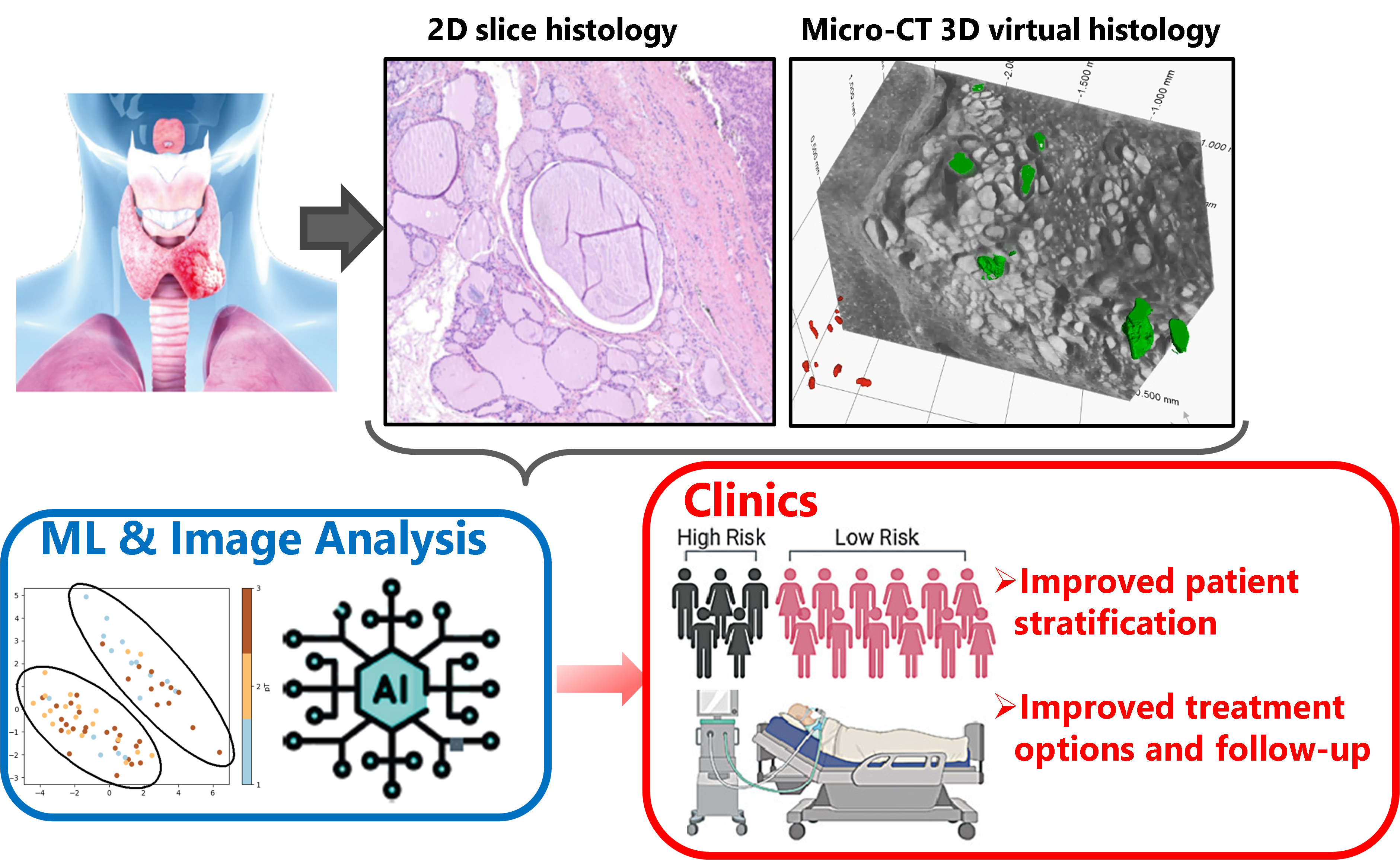
The diagnostic and prognostic evaluation of follicular thyroid nodules is complex, and conventional 2D slice histology suing H&E staining is inherently limited by sampling constraints. The reliance on morphological features like capsular and vascular invasion can sometimes lead to misdiagnosis, either underestimating or overestimating the severity of the condition. Consequently, unexpected tumor recurrence and overtreatment represent significant clinical challenges. To mitigate these limitations and enable precision medicine and diagnostics, emerging diagnostic approaches are exploring advanced imaging modalities. Whereas for the 3D imaging and ex-vivo evaluation of human tissues in biomedical and pathology applications of conventional clinical CT fails to produce sufficient contrast and visibility, X-ray phase-contrast micro-CT based 3D virtual histology enables non-destructive and comprehensive sampling of the entire tumor capsule in the formalin-fixed paraffin-embedded (FFPE) blocks. It brings clear potential clinical benefits as was shown by our research [1,2] furthermore it requires no further sample preparation of the FFPE blocks and preserves them for further analysis thus could be seamlessly integrated in the clinical workflow. These aspects render 3D digital histopathology by X-ray phase-contrast micro-CT to an extremely interesting tool to revolutionize research in tumor diagnostic for precision medicine. Our research extends to complementing and combining 3D X-ray histopathology with machine learning and data science methods as is depicted in the graphical abstract.

These are to help the pathologist's search for locating and identifying the above-mentioned tumor features (see image below) and in the decision making. In our present study we specifically focus on examining retrospective recurrence cases of thyroid tumors where we believe the largest clinical impact of our method can be martialized.
In order to address these questions, we work closely with clinicians and clinical researchers at University Hospital Bern, the Pathology Institute of the University Bern and several university hospitals of neighboring EU countries.

References
[1] Tajbakhsh, K., Stanowska, O., Neels, A., Perren, A., Zboray, R.: 3D virtual histopathology by phase-contrast X-ray micro-CT for follicular thyroid neoplasms, IEEE Trans Med Imaging. 2024 Mar 4; PP. doi: 10.1109/TMI.2024.3372602
[2] Tajbakhsh, K., Neels, A., Fadeeva, Larsson. J., Stanowska, O., Perren, A., Zboray, R.: A survey of micro-CT for 3D virtual histology of FFPE blocks, IEEE Access (2024)
PD Dr. Robert Zboray
Center for X-ray Analytics
Phone: +41 58 765 4602
We highly appreciate kind support of the Swiss Cancer Foundation, the Alfred und Annalise Sutter-Stöttner Stiftung, the stiftup, the Spendestiftungs Bank Vontobel, the Dr. Hans Altschüler Stiftung and the Conny-Maeva Charitable Foundation for our research.
